
Endobon® Bovine Xenograft
Particulate and Moldable
Sculpt with Precision Transition into Preservation.
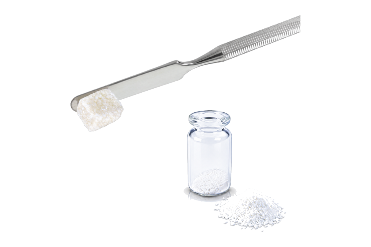
Sculpt with Precision Transition into Preservation
Shaping the future
Endobon is a bovine-derived cancellous xenograft designed for long-term space maintenance with optimal handling properties.1 The graft is available in two forms: particulate and moldable – giving clinicians flexibility to treat a range of defect types.
Endobon Bovine Xenograft Benefits
Shaping simplified
- Particles adhere together6 for seamless placement
- Moldable resists washout during irrigation3
Bovine-derived
- Sustains for long-term space maintenance delivering lasting structural support1
- Fully de-proteinized hydroxyapatite through controlled high-temperature processing2
- Histological data indicates Endobon functions as an effective osteoconductive scaffold, showing no significant difference as compared to conventional xenografts5
Osteoconductive scaffold
- Hydrophilicity2 promotes immediate capillary saturation 6,7
- Encourages osteoblast differentiation with clinically relevant bone formation3
- Hydrated matrix remains cohesive for predictable space maintenance6
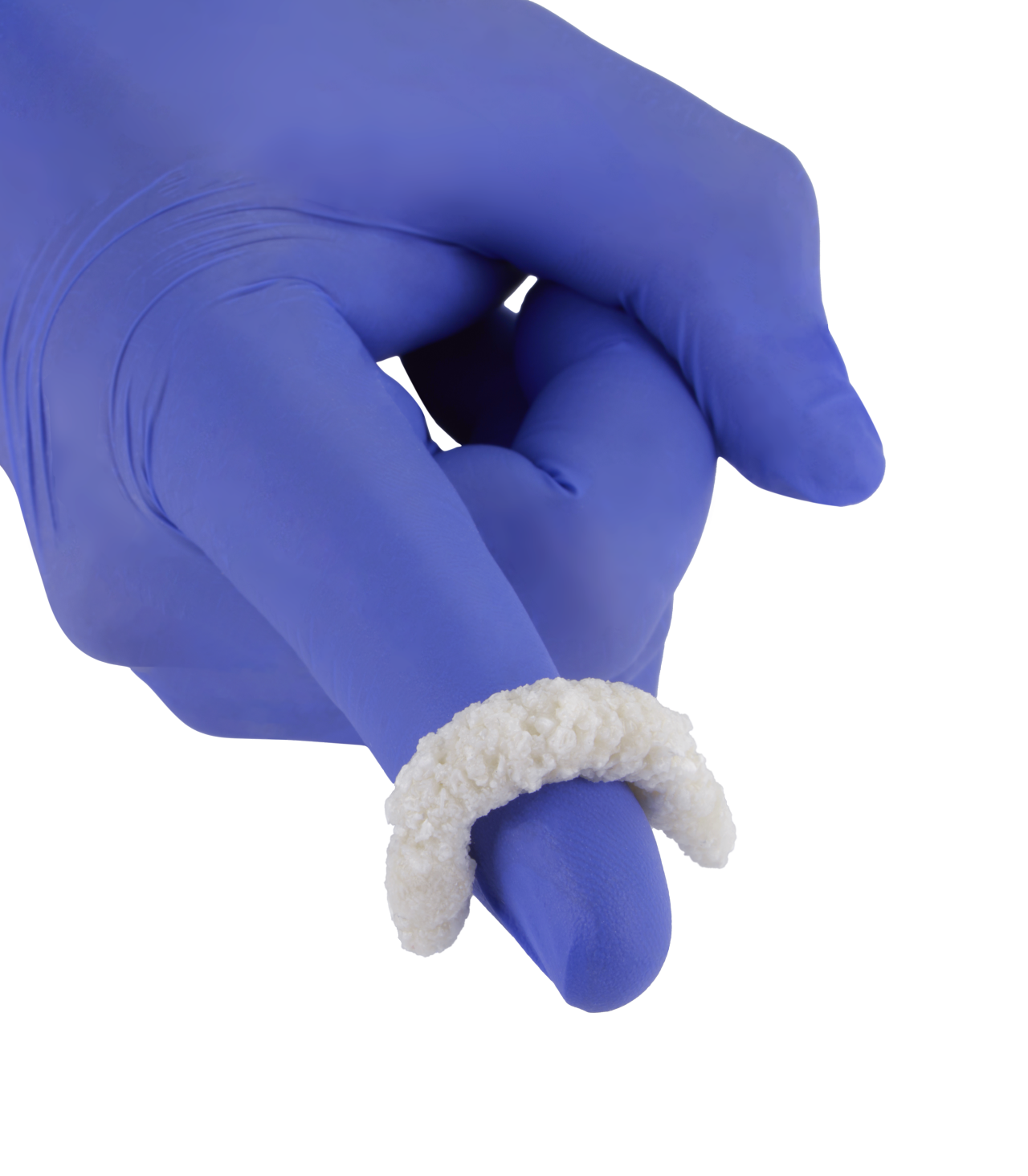
Endobon Moldable Features
Control where it counts
Shaping simplified
- Unique carrier3 facilitates conformable shaping properties
- Adapts to the geometry of the defect3,6
Hydrophilic advantage
- Carrier creates a wet-swell network environment that increases new bone formation5
- Carrier has a positive effect on osteoblast differentiation3,5
Stable adhesion
- Material adheres firmly to the surgical site, providing a stable scaffold for new bone formation3
- Resists irrigation and reduces scatter 3,6
Endobon Particulate Features
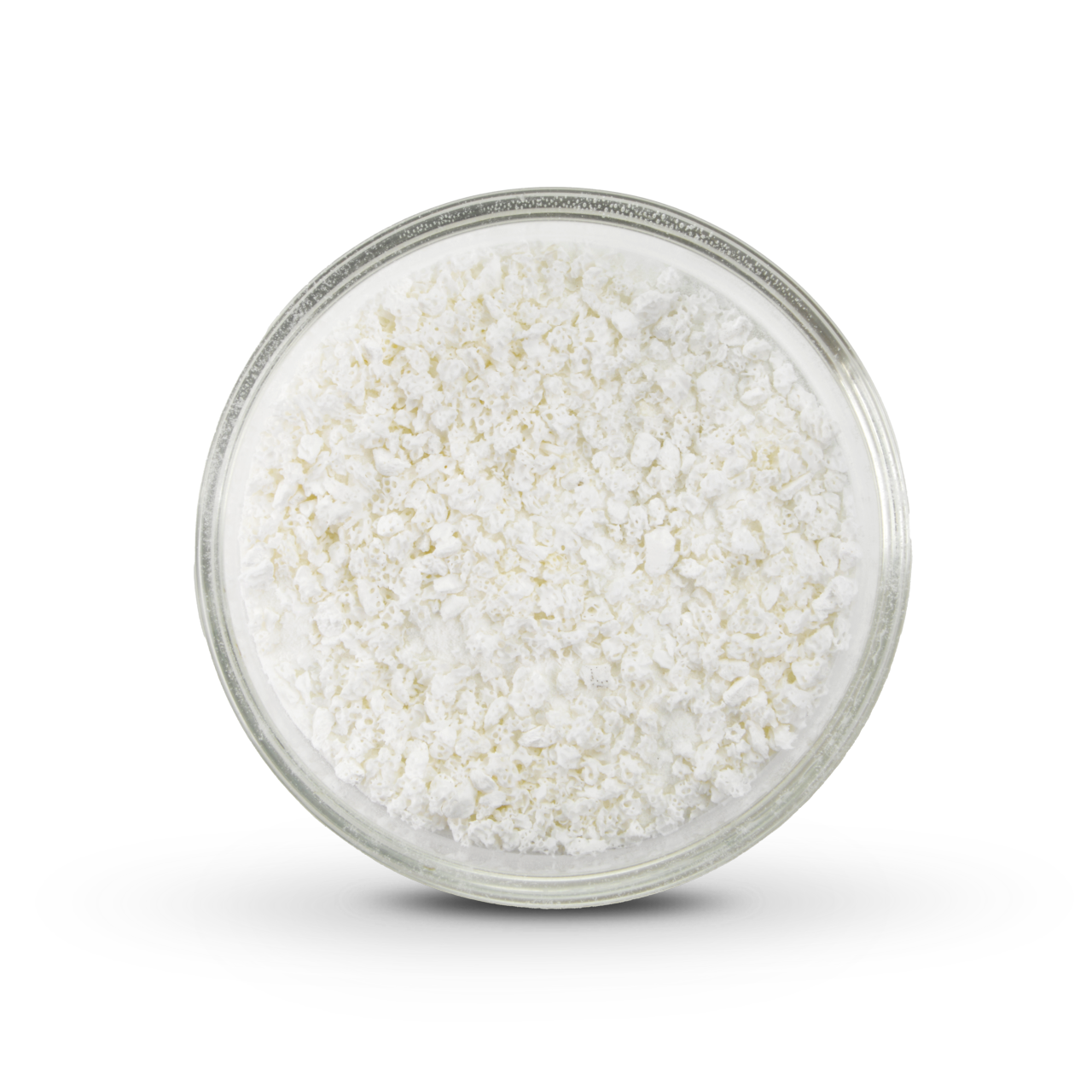
Particles with purpose
Volume stability
- Slow resorbing particles maintain volume during healing
- Facilitates the foundation for new bone formation 5
Hydrophilic cohesion
- Particulate hydrates rapidly 2
- Particulate adheres to one another for easy transfer to the defect site 3
Structural architecture
- Encourages an optimal environment for osteoblast adhesion and blood vessel formation 5
- 70% porosity maximizes surface area for cellular attachment 3
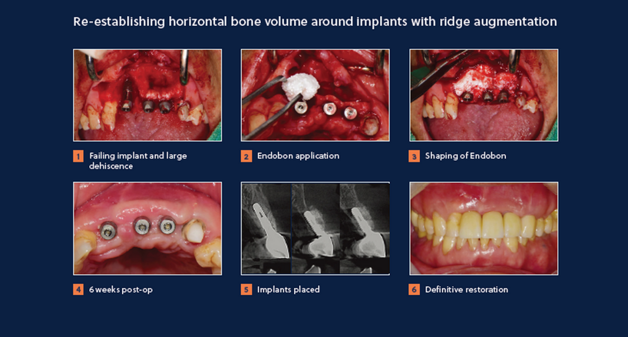
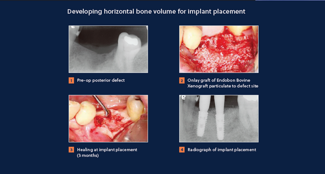
Clinical Cases
See the workflow in practice

Endobon Bovine Xenograft Resources
Videos
Three Simple Steps to Better Dental Care

Need Help? Contact Us
For any questions, concerns, or support, our dedicated team is here to assist you.

Explore Our Education
Learn more in our dental education platform for in-person or virtual programs.

Order Online With Ease
Use our online platform for quick order placement, returns, and complaints.
Additional Information
Product clearance and availability may be limited to certain countries/regions. Please contact your local sales representative or customer service team.